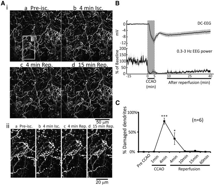
Figure 2.
Global ischemia induces dendritic damage to PV-neurons (inhibitory interneurons) which rapidly recovers following reperfusion. Ai, 2P imaging (0–60 μm in depth) following 5 min global ischemia and reperfusion. The white-boxed area on the top left corresponds to a region in higher magnification illustrations below (Aii). B, Changes of average DC potential and spontaneous EEG power (0.3–3 Hz) are shown (mean ± SEM, n = 6). C, Average percentage of damaged dendrites during global ischemia and reperfusion are shown (mean ± SEM, *p < 0.05, ***p < 0.001, one-way ANOVA followed by Bonferroni's post-test, compared with pre-CCAO group, n = 6). Isc, ischemia; Rep, reperfusion.

