
Figure 1.
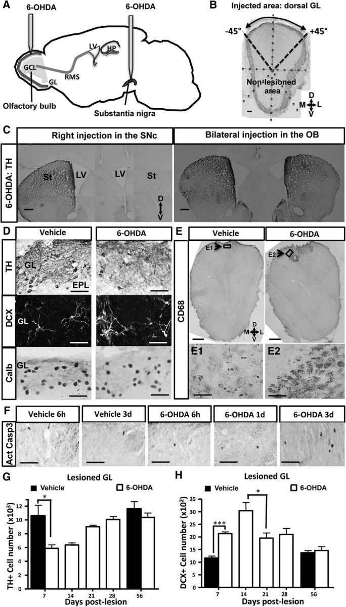
Transient dopamine cell loss after a 6-OHDA-induced lesion of the OB. A, Design of the experimental procedure. A 6-OHDA-containing solution, or vehicle alone, was infused into the right SN or into the dorsal part of each OB. B, TH immunostaining delineates the injected and noninjected areas in a coronal OB section. The image was divided into 8 sectors of 45°. C, TH immunoreactivity of coronal sections of striata and SVZ at 1 month after 6-OHDA injection into the right SN (left) or the two OBs (right). The efficacy of DA denervation is demonstrated by low levels of TH expression in the right striatum and right SVZ after neurotoxin injection into the right SN (left), depicting the extent and specificity of the lesion. Conversely, the right panel represents no alteration of TH expression in the two striata and SVZ after injection of 6-OHDA into the OB. D, Immunoreactivity for TH (top), DCX (middle), and Calb (bottom) of the lesioned GL at 7 d after injection into the OB of vehicle (left) or 6-OHDA (right). 6-OHDA reduces the number of DAergic cells expressing TH in the dorsal GL. Conversely, 6-OHDA increases DCX number but does not change Calb number in the same area. E, Immunoreactivity for CD68 (E1, E2, top) of the dorsal OB at 7 d after injection into the OB of vehicle (left) or 6-OHDA (right). 6-OHDA activates microglial cells as revealed by CD68 staining. Note the restricted presence of activated microglia in the dorsal part of the OB, depicting the extent and specificity of the lesion. Arrow indicates the area shown at higher magnification in E1 and E2. F, Time course of apoptotic cells expressing active cleaved casp 3 in the lesioned GL after 6 h, 1 d, and 3 d after 6-OHDA injection. The number of apoptotic cells is increased at day 3 after OB lesion. G, Time course of TH+ PGNs located in the lesioned GL after OB lesion (n = 4 or 5 animals per group). The number of TH-expressing neurons is transiently reduced in the dorsal GL after OB lesion. H, Time course of DCX-positive PGNs located in the lesioned GL after OB lesion (n = 4 or 5 per group). The number of developing neurons expressing DCX peaks at day 14 in the dorsal GL after OB lesion and resumes 21 d later. *p < 0.05. ***p < 0.001. Scale bars: B, C, 100 μm; D, 20 μm; E, 100 μm; E1, E2, 20 μm.