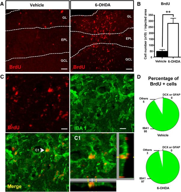
Figure 3.
Microglial cells, but not neuroblasts, proliferate in the 6-OHDA-lesioned OB. A, BrdU immunostaining in the OB of a vehicle (left) and a 6-OHDA-injected OB (right) at 7 d after lesion. BrdU-positive cells were restricted to the dorsal part of the OB, mainly in the GL, but also in the external plexiform layer and GCL. B, Quantification of BrdU-positive cells in the injected area (dorsal part of the OB) at 7 d after lesion (n = 4 mice per group). C, BrdU and IBA1 immunostaining in the GL of a 6-OHDA-injected OB at 7 d after lesion. BrdU was only found in microglial cells expressing IBA1. Arrow indicates the cell shown at higher magnification in C1. D, Percentage of BrdU-positive cells colabeled with one of three cell markers: GFAP for astrocytes, DCX for developing neurons, and IBA1 for active microglial cells. Cell counts were performed in the dorsal GL of vehicle and 6-OHDA-lesioned mice at 7 d after lesion (n = 4 mice per group, ∼100 cells analyzed per mouse). Nearly all fast-proliferating cells positive for BrdU coexpressed IBA1. **p < 0.01, compared with vehicle. Scale bars: A, 50 μm; C, 10 μm.

