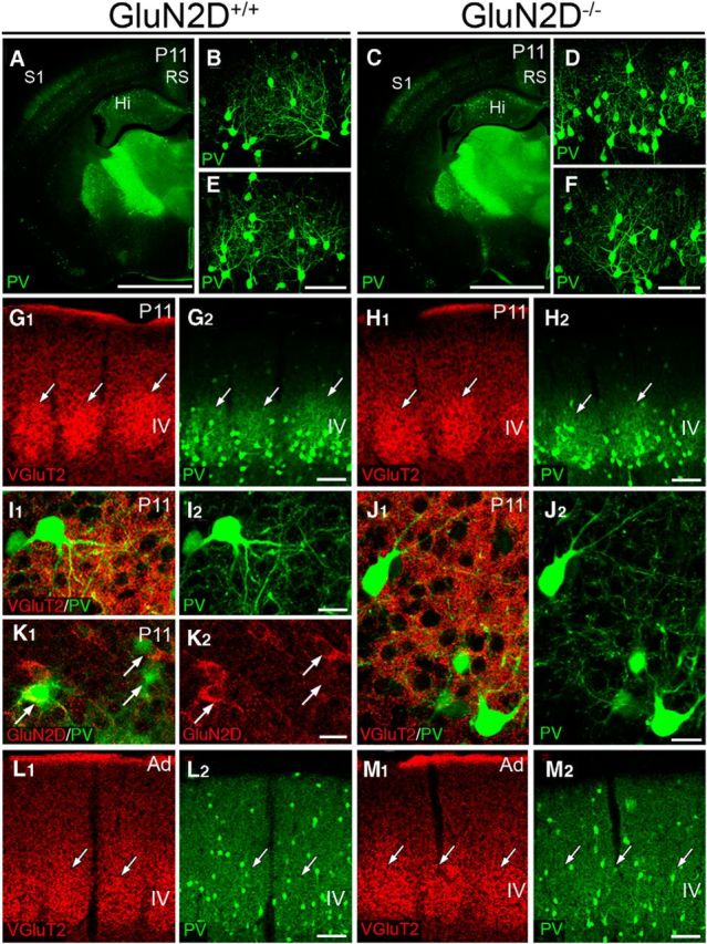
Figure 10.

Normal cytodifferentiation of PV-positive interneurons in the S1 of GluN2D−/− mice. A–F, PV immunofluorescence in GluN2D+/+ (A–C) and GluN2D−/− (D–F) mice at P11. PV expression started to increase in the somatosensory cortex (S1) and retrosplenial cortex (RS) (A, D), and whisker-related patterning by PV expression is evident (B, C, E, F) in both mice. G–J, Double immunofluorescence for VGluT2 (red) and PV (green) shows that whisker-related patterning by PV-positive interneurons matches with that by thalamocortical afferents (G, H). In both mice, cell bodies and dendrites of PV-positive interneurons similarly form baskets surrounding barrel hollows, in which VGuT2(+) thalamocortical terminals are densely distributed (I, J). K, Double immunofluorescence for GluN2D (red) and PV (green) in GluN2D+/+ mice at P11. At this stage, PV is upregulated in some interneurons, including those expressing GluN2D (arrows). L, M, Whisker-related patterning by PV-positive interneurons is no longer seen in adult GluN2D+/+ (L) and GluN2D−/− mice (M). Scale bars: A, D, 1 mm; B, C, E, F (in C, F), 200 μm; G, H, L, M, 100 μm; I–K, 20 μm.
