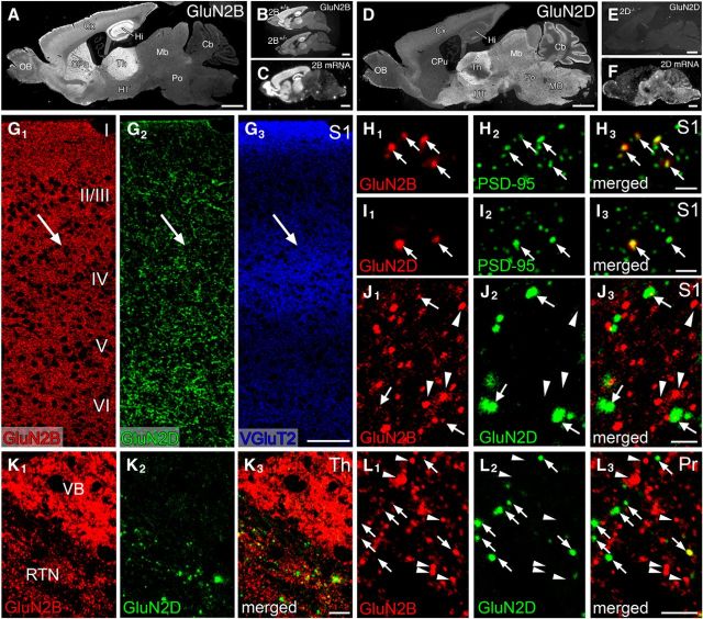
Figure 7.
Immunofluorescence showing distinct distribution pattern of GluN2B and GluN2D immunoreactivities at trigeminal relay stations of adult mice. A, D, Overall staining patterns in wild-type mouse brains for GluN2B (A) and GluN2D (D). B, E, The specificity is indicated by substantial reduction of GluN2B immunolabeling in GluN2B+/− brain compared with GluN2B+/+ brain (B), and by the lack of GluN2D immunolabeling in GluN2D−/− brain (E). C, F, Isotopic in situ hybridization for GluN2B (C) and GluN2D (F) mRNAs in adult mouse brains. G, Triple immunofluorescence for GluN2B (red), GluN2D (green), and VGluT2 (blue) in the S1. Arrows indicate the position of barrels labeled for VGluT2. H, I, High-power views of double immunofluorescence for PSD-95 (green) and GluN2B (red) or GluN2D (red) in the S1. Punctate labeling for both subunits (arrows) well overlaps with PSD-95 signals. J–L, High-power views of double immunofluorescence for GluN2B (red) and GluN2D (green) in the S1 (J), VB and RTN (K), and Pr (L). Intense GluN2B-positive puncta virtually lack GluN2D signals (arrowheads), and GluN2D puncta often possess weak GluN2B signals (arrows). I-VI, Cortical layers I through VI; Cb, cerebellum; CPu, caudate-putamen; Cx, cortex; Hi, hippocampus; HT, hypothalamus; Mb, midbrain; MO, medulla oblongata; OB, olfactory bulb; Po, pons; Th, thalamus. Scale bars: A–F, 1 mm; G, 100 μm; H–J, 2 μm; K, 10 μm; L, 5 μm.

