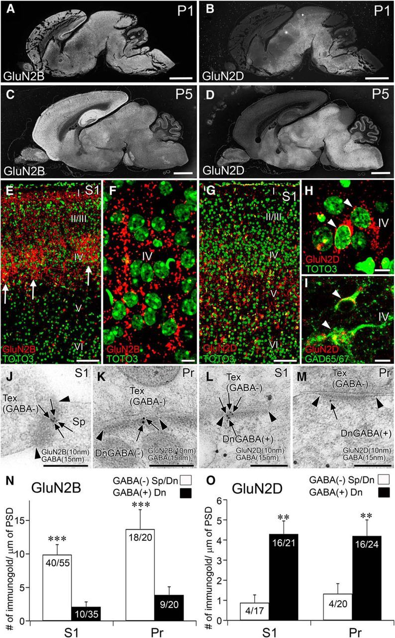
Figure 9.

Segregated synaptic expression of GluN2B and GluN2D is preserved at neonatal trigeminal relay stations. A–D, GluN2B (A, C) and GluN2D (B, D) immunolabeling in the brain of wild-type mice at P1 (A, B) and P5 (C, D). E, F, GluN2B immunofluorescence (red) and nuclear counterstaining with TOTO-3 (green) in the S1. Note that barrel hollows (arrows) are filled with intense GluN2B(+) puncta. G–I, GluN2D immunofluorescence (red) with nuclear counterstaining with TOTO-3 (green, G, H) or with GAD65/67 immunofluorescence (green, I) in the S1. Note accumulation of GluN2D in perikarya of GAD65/67(+) neurons (I, arrowheads). J–M, Double-labeling postembedding immunogold for GluN2B (J, K) and GluN2D (L, M) in the S1 (J, L) and Pr (K, M). N, O, Summary bar graphs representing preferential expression of GluN2B (N) and GluN2D (O) at synapses on GABA(−) and GABA(+) postsynaptic compartments, respectively, in the S1 and Pr. **p < 0.01 (U test). ***p < 0.001 (U test). Scale bars: A-D, 1 mm; E, G, 100 μm; F, H, I, 10 μm; J–M, 200 nm.
