Figure 1.
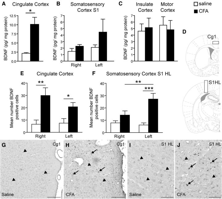
Peripheral inflammation induced an increased BDNF expression in both the cingulate and primary sensory cortices. A–C, ELISA titration showing that BDNF concentration was increased markedly in the cingulate cortex (pool of bilateral sites; A) and to a lesser extent in the primary sensory cortex (B) and unchanged in the insulate and motor cortices (C) 24 h after the induction of peripheral inflammation by unilateral intraplantar injection of CFA. E–J, Immunohistochemical staining of BDNF (G–J) and its quantification (E, F) reveals an increased number of BDNF-positive cells in the right and left cingulate cortex (E) in inflamed animals. There is also an increase in the contralateral somatosensory cortex, hindlimb part (F). D, Schematic showing the area used to quantify the number of BDNF-positive cells in the cingulate cortex (D, top) and the S1HL (D, bottom). G, H, Representative BDNF immunostaining in the ACC of a control (G) and a CFA-injected rat (H). I, J, Representative BDNF immunostaining in the S1HL of a control (I) and a CFA-injected rat (J). Scale bars: G, H, 200 μm; I, J, 100 μm. All data are expressed as mean ± SEM; N = 5 CFA-treated animals, n = 5 control animals. Two-way ANOVA followed by Student-Newman–Keuls comparison post hoc test. B: F(1,18) = 2017, p = 0.176; E: F(1,25) = 16.62, p < 0.001; F: F(1,25) = 17.25, p < 0.001; interaction: F(1,25) = 4.95, p = 0.037; *p < 0.05, **p < 0.01, ***p < 0.001 versus control rats.

