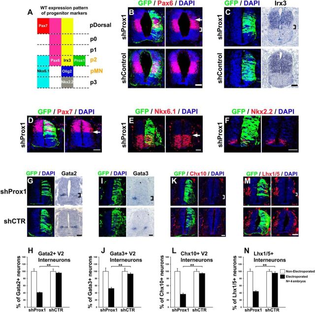
Figure 12.
Prox1 expression in p2 domain is required for the proper acquisition of p2 progenitor identity and V2 interneuron generation. A, Schematic diagram of the wild-type expression pattern of progenitor markers in ventral spinal cord. The p2 and pMN are marked with orange color to indicate that the boundary between these domains is affected by shProx1. B, Double GFP/Pax6 immunostainings in cryosections 48 h a.e. with shProx1 (top) or shControl (bottom). The square bracket indicates the contraction of ventral boundary of Pax6+ domain in the shProx1 electroporated side. Conversely, the arrow indicates the dorsal boundary of Pax6+ domain, which remains unaffected under the same conditions. C, GFP/DAPI staining and in situ hybridization for Irx3 in consecutive sections 48 h a.e. with shProx1 (top) or shControl (bottom). The square bracket indicates the contraction of ventral boundary of Irx3+ domain. D, Double GFP/Pax7 immunostaining in cryosection 48 h a.e. with shProx1. The arrow indicates the ventral boundary of Pax7+ cells, which remains unaffected. E, Double GFP/Nkx6.1 immunostaining in cryosection 48 h a.e. with shProx1. The arrow indicates the dorsal boundary of Nkx6.1+ cells, which remains unaffected. F, Double GFP/Nkx2.2 immunostaining in cryosection 48 h a.e. with shProx1. Note that the domain of Nkx2.2+ cells (p3) is not affected by shProx1. G, GFP/DAPI staining and in situ hybridization for Gata2 in consecutive sections 48 h a.e. with shProx1 (top) or shControl (bottom). H, Quantitative analysis of the Gata2+ area using ImageJ. For shProx1 versus shControl, **p < 0.01 (t test), n = 4 embryos. All cases referred to the electroporated side. I, GFP/DAPI staining and in situ hybridization for Gata3 in consecutive sections 48 h a.e. with shProx1 (top) or shControl (bottom). J, Quantitative analysis of the Gata3+ area using ImageJ. For shProx1 versus shControl, **p < 0.01 (t test), n = 4 embryos. All cases referred to the electroporated side. K, M, Double GFP/Chx10 (K) and GFP/Lhx1/5 (M) immunostainings in cryosections 48 h a.e. with shProx1 (top) or shControl (bottom). L, N, Quantitative analysis of the number of Chx10+ (L) and Lhx1/5+ cells (N). For Chx10+ cells: shProx1 versus shControl, **p < 0.01 (t test), n = 4 embryos; for Lhx1/5+ cells: shProx1 versus shControl, **p < 0.01 (t test), n = 4 embryos. All cases referred to the electroporated side. The square brackets indicate the domains of Gata2+ (G), Gata3+ (I), and Chx10+ (K) V2 interneurons, as well as Lhx1/5+ V2 interneurons (M), which are strongly reduced after shProx1 electroporation. Scale bars: 50 μm.

