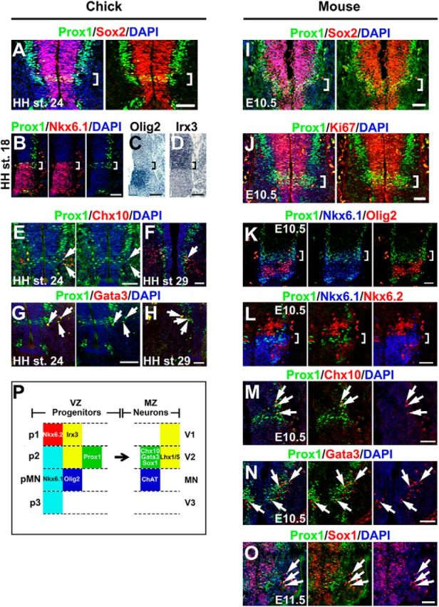
Figure 3.

Prox1 is expressed in the V2 interneuron progenitors in embryonic chick and mouse spinal cord. A, Double immunostaining of Prox1 (green) and Sox2 (red) in HH stage 24 chick spinal cord. The square brackets indicate the expression of Prox1 in the proliferating cells of the VZ, Prox1+/Sox2+ cells, exactly above the MN domain. B–D, Double immunostaining of Prox1 (green) and Nkx6.1 (red; B) and in situ hybridizations for Olig2 (C) and Irx3 (D) on adjacent cryosections from HH stage 18 chick spinal cord. The square brackets indicate the Prox1+ cells in p2 domain (Nkx6.1+, Olig2−, and Irx3+). E–H, Double immunostainings of Prox1 (green) and Chx10 (red; E, F) or Gata3 (red; G, H) in HH stages 24 (E, G) and 29 (F, H) chick spinal cord. The arrows indicate cells that coexpress Prox1 and the V2a interneuron marker, Chx10, or the V2b marker, Gata3. The partial colocalization could be explained by the previous observations that Prox1 expression is downregulated before acquisition of terminal neuronal identity in spinal cord interneurons (Misra et al., 2008; Kaltezioti et al., 2010). I, Double immunostaining of Prox1 (green) and Sox2 (red) in E10.5 mouse spinal cord. The square brackets indicate the expression of Prox1 in the proliferating cells of the VZ, Prox1+/Sox2+ cells, exactly above the MN domain. J, Double immunostaining of Prox1 (green) and Ki67 (red) in E10.5 mouse spinal cord. The square brackets indicate the expression of Prox1 in the proliferating cells of the VZ, Prox1+/Ki67+ cells, exactly above the MN domain. K, Triple Prox1 (green), Nkx6.1 (blue), and Olig2 (red) immunostaining in E10.5 mouse spinal cord. The square brackets indicate the Prox1+ cells in p2 domain (Nkx6.1+ and Olig2−). L, Triple Prox1 (green), Nkx6.1 (blue), and Nkx6.2 (red) immunostaining in E10.5 mouse spinal cord. The square brackets indicate the Prox1+ cells in p2 domain (Nkx6.1+ and Nkx6.2−). M–O, Double immunostaining of Prox1 and Chx10 (M), Gata3 (N), or Sox1 (O) in E10.5 or E11.5 mouse spinal cords, as indicated. The arrows indicate the cells that coexpress Prox1 and Chx10 (V2a), Gata3 (V2b), and Sox1 (V2c). The partial colocalization could be explained by the previous observations that Prox1 expression is downregulated before acquisition of terminal neuronal identity in spinal cord interneurons (Misra et al., 2008; Kaltezioti et al., 2010). P, In the schematic diagram, we summarize the experimental data showing that Prox1 is expressed in the p2 domain of VZ. Scale bars: 50 μm.
