Figure 4.

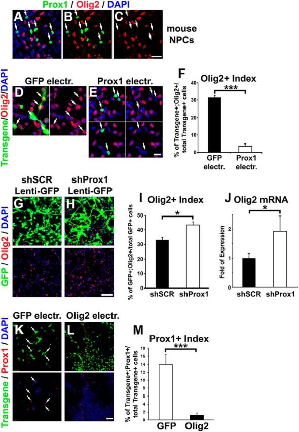
Prox1 is sufficient and necessary for proper regulation of Olig2 expression in NPCs derived from mouse spinal cord. A–C, Double Prox1 (green) and Olig2 (red) immunostaining of NPCs isolated from E12.5 mouse spinal cord and cultured in vitro. The Prox1+/Olig2− cells are indicated with arrows. D, E, Double GFP/Olig2 (D) and Flag/Olig2 (E) immunostainings of mouse NPCs electroporated with GFP or Prox1, respectively. Prox1 was detected with the anti-Flag antibody. Arrows indicate the transgene-positive cells. F, Quantification of the transgene+ cells that are Olig2+ (percentage of transgene+;Olig2+/total transgene+). *** p < 0.001 (t test), n = 5. G, H, Double GFP/Olig2 immunostainings of mouse NPCs infected with lentiviruses overexpressing GFP and either scramble shRNA (shSCR, G) or shRNA targeting murine Prox1 (shProx1, H). The efficiency of shProx1 vector has been previously shown (Foskolou et al., 2013). I, Quantification of the GFP+ cells that are Olig2+ in NPCs infected with GFP-shSCR or GFP-shProx1 lentiviruses, as indicated (percentage of GFP+;Olig2+/total GFP+). *p < 0.05 (t test), n = 5. J, Real-time RT-PCR analysis for Olig2 gene expression in mouse NPCs infected with GFP-shSCR or GFP-shProx1 lentiviruses, as indicated, *p < 0.05 (t test), n = 5. K, L, Double GFP/Prox1 (K) or myc/Prox1 (L) immunostainings of mouse NPCs electroporated with GFP or Olig2 (carrying the myc tag), respectively. Double-positive cells are indicated with the white arrows. M, Quantification of transgene+ cells that are Prox1+ (percentage of transgene+;Prox1+/total transgene+) in NPCs electroporated with GFP or Olig2. ***p < 0.001 (t test), n = 4. Scale bars: A–C, 20 μm; D, E, 10 μm; G, H, K, L, 50 μm.
