Figure 7.
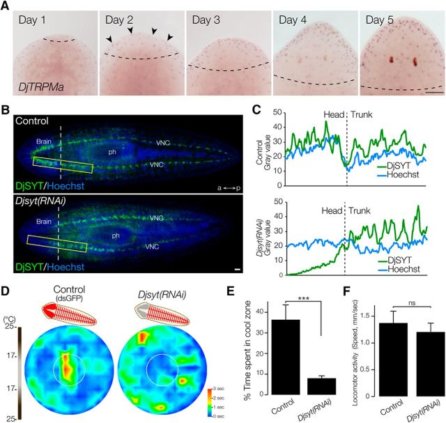
Brain neural activity is required for thermotaxis. A, Expression pattern of DjTRPMa during head regeneration. The dashed line indicates the border between the newly formed region and old stump region. Arrowheads indicate newly regenerated DjTRPMa-expressing cells. Scale bar, 300 μm. B, Readyknock of Djsyt. Left, Immunohistochemical detection of DjSYT protein shown in green in control (top) and in Readyknock (bottom) animals 7 d after decapitation. Samples were stained with Hoechst 33342 (for nuclei, shown in blue) to visualize planarian tissues, including brain. The dashed line indicates the border between the newly formed region and old stump region. ph, pharynx; a, anterior; p, posterior. Scale bar, 200 μm. C, Graphs show fluorescence intensity of DjSYT and Hoechst staining corresponding to yellow-boxed areas in B. D, Heat map of control and Djsyt(RNAi) planarians at 7 d of head regeneration using a thermal gradient dish (n = 10, t = 180 s). E, The time spent in the cool target area is shown as mean ± SEM of 10 independent animals. Synaptic transmission in brain-defective planarians produced by Djsyt(RNAi) did not result in normal thermotaxis. ***p < 0.005. F, Locomotor activity was not affected by Djsyt(RNAi). ns, Not significantly different.

