Figure 3.

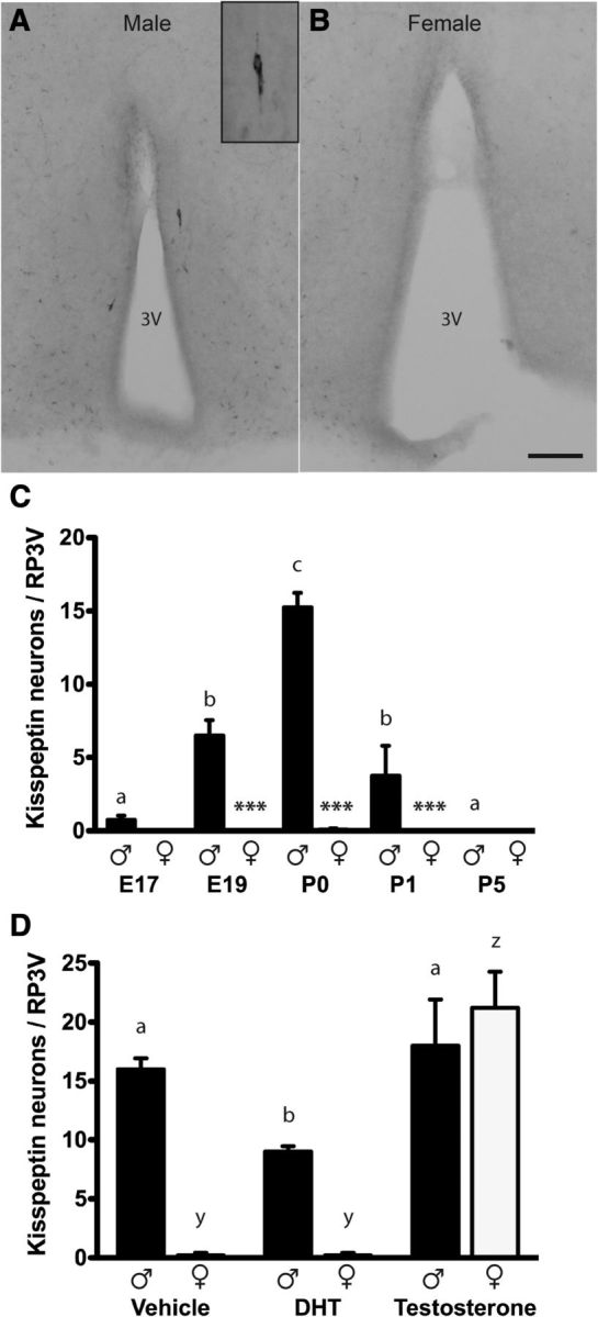
Sexually dimorphic expression of RP3V kisspeptin neurons in perinatal mice is regulated by prenatal testosterone exposure. Kisspeptin-immunoreactive cell bodies were detected in the RP3V of P0 male (A) but not female (B) mice. C, Quantitative analyses (mean ± SEM) of the number of kisspeptin-immunoreactive neurons in the RP3V in male and female mice from E17 to P5 (n = 2–12/sex/age). D, Quantitative analyses (mean ± SEM) of the number of kisspeptin-immunoreactive neurons in the RP3V in P0 male and female mouse pups from dams treated with vehicle, or 1 mg of either DHT or testosterone on E18 (n = 4–5/sex/treatment). Inset shows a higher power image of a single kisspeptin neuron. Scale bar, 100 μm. Groups were compared with a two-way ANOVA for sex and age/treatment with post hoc Student–Newman–Keuls pairwise comparisons. C, Sex (p < 0.001), age (p < 0.001), and interaction (p < 0.001); asterisks indicate differences between males and females at the ages indicated ***p < 0.001. D, Sex (p < 0.001), treatment (p < 0.001), and interaction (p < 0.001). Bars labeled with different letters are significantly different from each other at either p < 0.05 or p < 0.001 (see text). 3V, third ventricle.
