
Figure 6.
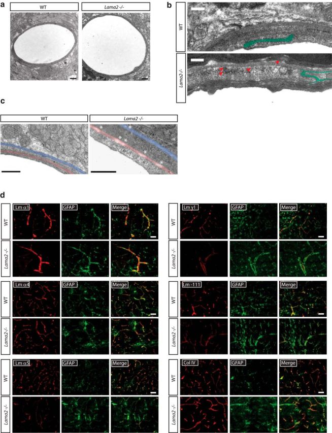
Altered structure and composition of the BBB BL in Lama2−/− mice. a–c, Transmission electron micrographs to evaluate the ultrastructure of the gliovascular interface in Lama2−/− and WT littermate brains at postnatal day 21. a, No obvious defects were apparent in the brain capillary BLs of Lama2−/− mice. b, Tight junction morphology (green) in Additionally, endocytic vesicles (red arrows) were observed in Lama2−/− vascular endothelial cells. c, Regions of the parenchymal BL (blue) and endothelial BL (red) are discontinuous (white asterisk) in large-caliber blood vessels of Lama2−/− mice. d, Immunohistochemistry to detect components of the BL in conjunction with GFAP in the cerebral cortices of Lama2−/− and WT mice at postnatal day 21. Laminin α1, α4, α5, γ1, laminin-111 (a polyclonal antibody that recognizes laminin α1, β1, and γ1 subunits), and collagen IV (col IV) were detected in the gliovascular BL of Lama2−/− and WT brains. Laminin α5 immunoreactivity was reduced in Lama2−/− mice. Scale bars: a, 500 nm; b, 100 nm; c, 500 nm; d, 50 μm.