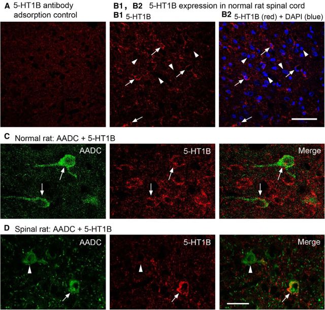
Figure 10.
Confocal images showing 5-HT1B receptors were expressed in the spinal cords of both normal and spinalized rats. A, When 5-HT1B antibody was adsorbed with the corresponding antigen peptide (100 μg/ml antibody solution), specific immunolabeling was very weak or nonexistent. B1, B2, Images from a region in the intermediate zone of normal rat spinal cord show that 5-HT1B receptors were expressed widely in the gray matter. 5-HT1B immunoreactivity was located in the cytoplasm and most likely also in the cell membrane (arrows). Arrowheads in B1 and B2 indicate several DAPI-labeled cells that were not 5-HT1B immunopositive. C, D, Images showing that 5-HT1B receptors were expressed in spinal AADC cells (arrows) in a normal (C) and a 75 day spinalized rat (D; arrows). Note that, in the spinalized rat, an AADC cell was 5-HT1B negative (arrowhead). Scale bars: (in A2) A–B2, 50 μm; (in D) C, D, 25 μm.

