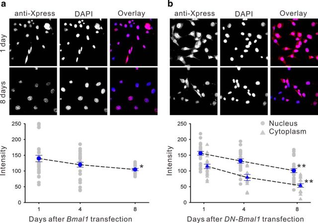
Figure 3.
Intracellular localization of wild-type mouse BMAL1 or DN-BMAL1 overexpressed in NIH 3T3 fibroblasts visualized by epitope tag immunostaining (anti-Xpress). a, Top, Exogenous BMAL1 was located in NIH 3T3 fibroblast nuclei. Bottom, The mean exogenous BMAL1 expression declined over 8 d in culture (blue data points indicate means ± SE relative to the intensity of DAPI nuclear staining; n ≥ 6; gray data points indicate individual measurements;*p < 0.05 by one-way ANOVA). b, DN-BMAL1 was distributed in the nucleus and cytoplasm, and also declined over 8 d in culture; **p < 0.01 by one-way ANOVA.

