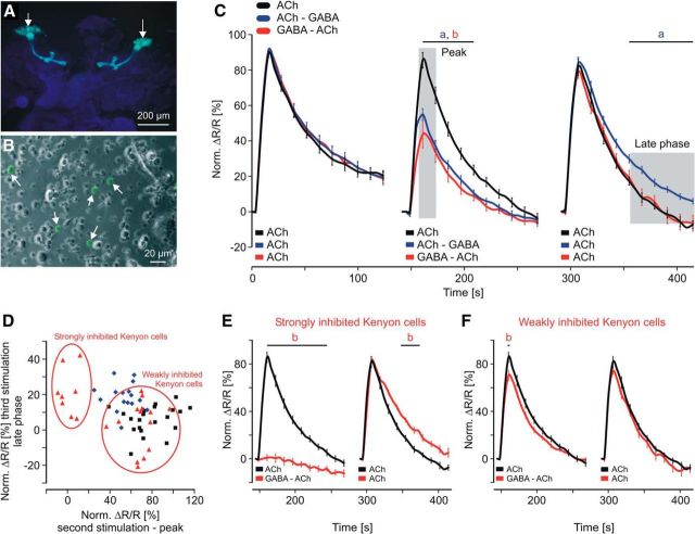
Figure 4.
Calcium responses in a defined subset of isolated Drosophila KCs during temporal pairing of ACh and GABA. A, Fluorescence image of larval brain whole mount (201y). The calcium-sensitive fluorescence protein cameleon is specifically expressed in a subset of MB KCs involved in learning. B, The isolated KCs were identified by their characteristic cameleon fluorescence (arrows). C, Pairing of ACh and GABA during the second stimulation significantly inhibited the calcium response. ACh–GABA pairing led to elevated calcium levels during the late phase of the third stimulation. Shaded areas indicate averaged time windows used in D. Data derived from at least five independent preparations (ACh: n = 22 neurons; ACh–GABA: n = 17 neurons; GABA–ACh: n = 22 neurons). Two-way ANOVA followed by Bonferroni post hoc test: a, ACh vs ACh–GABA; b, ACh vs GABA–ACh; p < 0.02. D, Calcium responses for single KCs showed substantial pairing-specific differences and division into subpopulations with different properties. E, F, Strongly inhibited KCs showed no calcium response during the second stimulation and elevated calcium levels during the late phase of the third stimulation. The other subpopulation of KCs was only weakly inhibited and showed no difference during the third stimulation. Two-way ANOVA followed by Bonferroni post hoc test: b, ACh vs GABA–ACh; p < 0.02.

