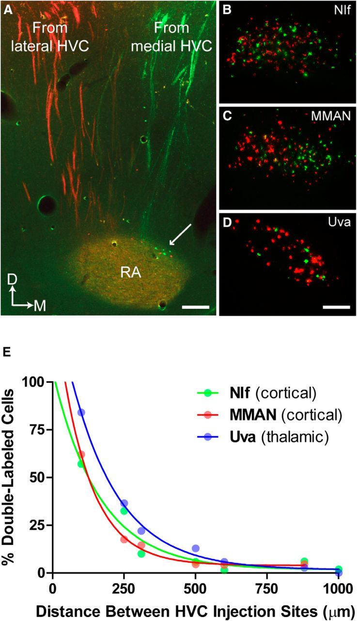
Figure 9.

Parallel organization of HVC extrinsic connectivity. Tracer injections in medial (DiO, green) and lateral (DiI, red) HVC show that the extrinsic connectivity of HVC supports a partitioning of vocal function across medial and lateral portions of HVC. Images show that medial and lateral HVC send axons to RA in distinct pathways (A) and receive distinct sources of afferent input from NIf, MMAN, and UVA (B–D). This pattern of anterograde and retrograde labeling was observed in all birds (N = 4) that received dye injections into medial and lateral HVC. A, Arrow indicates the small population of neurons in dorsal RA that are reciprocally connected to HVC (Roberts et al., 2008). These cells were also found to project medial HVC (green) or lateral HVC (red), but not both. Scale bars: A, 165 μm; D, 120 μm. E, Demonstration of experimental control over tracer labeling. Double-labeling of neurons in HVC afferent nuclei (NIf, MMAN, Uva) is an exponential function of the medial–lateral distance between ≤40 nl DiI and DiO injections in HVC. Data are single hemisphere values from N = 7 birds. The exponential function indicates that axon terminals from NIf, MMAN, and Uva neurons are narrowly targeted to either medial or lateral HVC.
