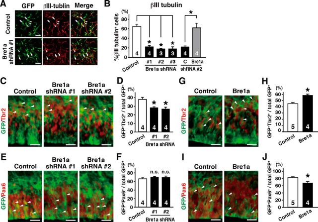
Figure 8.
Bre1a facilitates the differentiation of neural precursor cells. A, B, Primary neuroepithelial cells from E10.5 forebrains were transfected with Bre1a or control shRNAs and GFP expression plasmids, and cultured in differentiation condition for 3 d. Bre1a or control expression plasmids were cotransfected with Bre1a-shRNA 2 for rescue experiments. A, Cells were immunostained for GFP and βIII tubulin. Arrowheads and arrows indicate GFP+/βIII tubulin+ and GFP+/βIII tubulin− cells, respectively. B, Quantification of GFP+/βIII tubulin+ cells. C–J, Bre1a or control shRNA, pCX-Bre1a, or control expression plasmids were microinjected into the lateral ventricle of E13.5 embryos and electroporated into the dorsal forebrain in utero. The embryos were fixed 24 h after electroporation, and the brains were analyzed. C–J, Cryosections were immunostained for GFP and Tbr2 (C, G) or Pax6 (E, I). GFP+/Tbr2+ (D, H) or GFP+/Pax6+ (F, J) cells were counted. Scale bars: A, 50 μm; C, E, G, I, 100 μm. Error bars indicate SEM, and n values are shown in columns. *p < 0.05 by one-way ANOVA followed by Dunnett's post hoc comparison (B, D, F) or by Student's t test (H, J).

