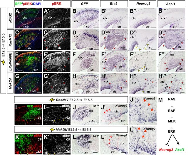
Figure 3.
RAS functions through the ERK branch to regulate the Neurog2-Ascl1 genetic switch in cortical progenitors. E12.5→E15.5 electroporations of a pCIG2 control vector (A, A′, B–B‴), RasV12 (C, C′, D–D‴), bRafV600E (E, E′, F–F‴), MekCA (G, G′, H–H‴), RasN17 (I, I′, J–J″), or MekDN (K, K′, L–L″) vectors (expressing GFP). Transfected brains were analyzed for coexpression of GFP with pErk (A, A′, C, C′, E, E′, G, G′, I, I′, K, K′), or for transcripts for GFP (B, D, F, H, J, L), Etv5 (B′, D′, F′, H′), Neurog2 (B″, D″, F″, H″, J′, J″, L′, L″), and Ascl1 (B‴, D‴, F‴, H‴). Dashed lines outline the transfected region in the neocortex. Red arrowheads mark ectopic pERK (C′, E′, G′), Etv5 (D′, F′, H′), Neurog2 (H″, J′, J″, L′, L″), and Ascl1 expression (D‴, F‴, H‴), whereas yellow arrowheads mark transfected areas in which Neurog2 (D″, F″, H″) or pERK (I, I′, K, K′) expression was extinguished. M, Schematic illustration of repression of Neurog2 expression and induction of Ascl1 expression by RAS/ERK signaling. Scale bars: 250 μm. CP, cortical plate; ctx, neocortex; str, striatum.

