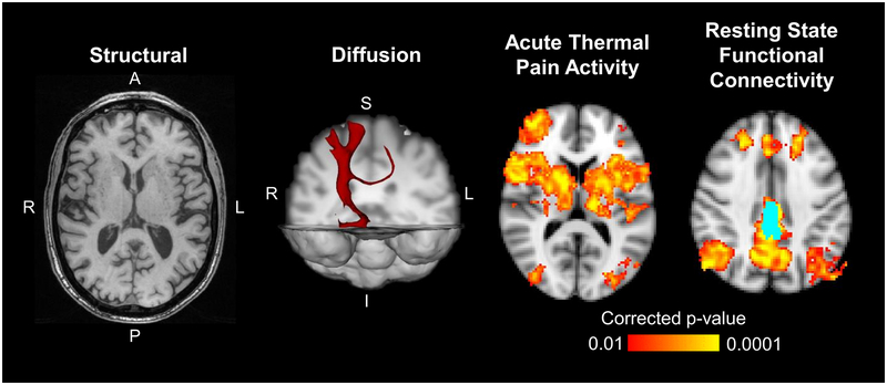
Figure 5-.
Example structural, diffusion, and functional brain images are shown. The axial structural image was acquired using 3D MPRAGE T1-weighted gradient-echo sequence and can provide morphometric properties of the gray matter. The diffusion example shows a 3D tractography map using the right ventral posterolateral nucleus of the thalamus as a seed. The axial functional images show average group activation from an acute thermal pain stimulus applied to the lower back and group average connectivity to the bilateral posterior cingulate cortices (light blue).

