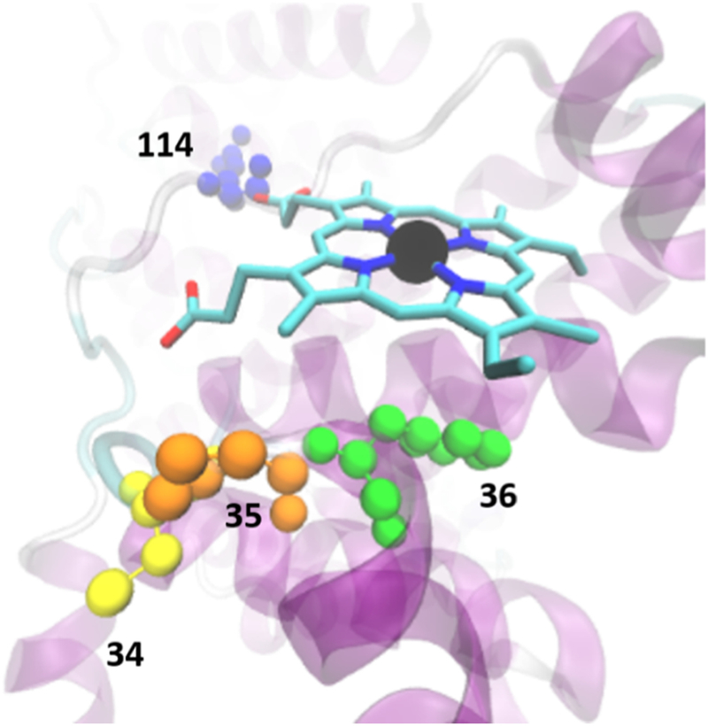
Figure 7.

Representative lowest energy binding configuration for Mn(III)PPIX and HSA at the Cys34 (yellow) location. The figure shows the contacts of the porphyrins with Pro35 (orange) and Arg114 (blue) as well as the aromatic interaction with Phe36 (green).
