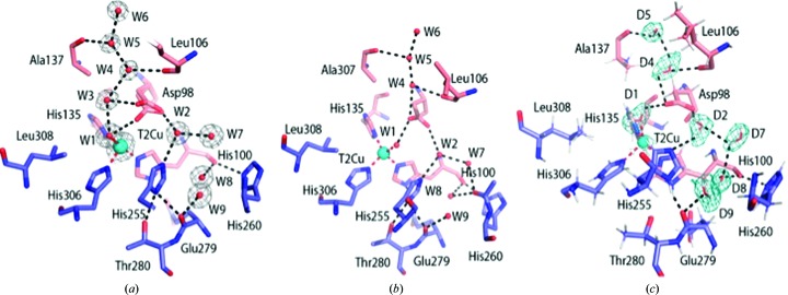
Figure 2.
Water structure in the catalytic pocket and substrate-entry channel. (a) In the structure of oxidized AcNiR determined by SF-ROX, Asp98 (AspCAT) has a dual conformation; the usual proximal conformation hydrogen-bonds to two waters (W3 and W4), while the distorted proximal position, which is observed for the first time, hydrogen-bonds directly to the T2Cu water ligand. The waters (W3 and W4) are part of the ordered water network in the substrate-entry channel. Both proximal conformations of AspCAT are hydrogen-bonded via Oδ1, with the water W2 linking His255 (HisCAT) to AspCAT. 2F o − F c electron density is contoured at the 1σ level and is shown as a grey mesh. (b) In the atomic resolution crystal structure (PDB entry 2bw4) the proximal conformation is hydrogen-bonded to the ligated water W1A with an occupancy of 0.8. Water W1B with an occupancy of 0.2 is not shown for simplicity. (c) In the neutronOX structure, AspCAT is in a single proximal conformation. The 2F o − F c nuclear scattering-length density map is contoured around selected heavy waters at the 1σ level and is shown as a teal mesh. Atoms are coloured by element, with different colour schemes used for the different chains. The T2Cu is shown as a cyan sphere, D2O water molecules are shown as red and white sticks and water molecules are shown as small red spheres. Metal-coordinating bonds are shown as red dotted lines. Selected hydrogen bonds are shown as black dotted lines.

