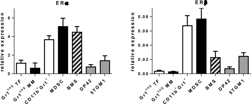
Figure 2. Expression of ERs in myeloma and BM cells.

Expression of ERα (left panel) and ERβ (right panel) was detected by RT-PCR in Gr1neg BM cells isolated from tumor-free (TB) or MM-bearing (MM) mice, CD11b+Gr1+ cells isolated from BM of TF mice, MDSC from BM of MM-bearing mice, BMS established in vitro, and indicated MM cell lines. Note the scale difference between the panels.
