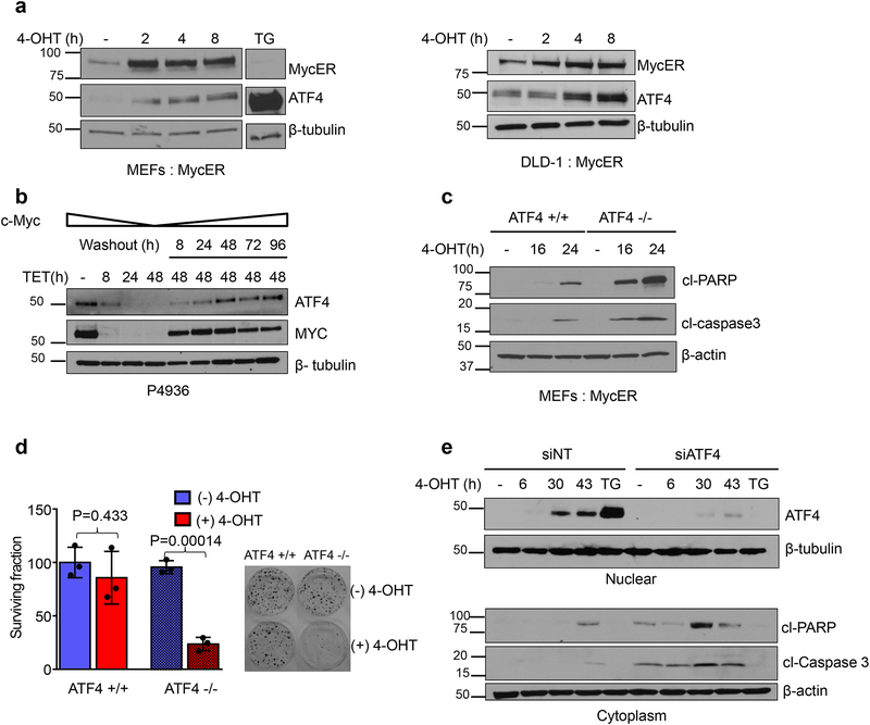
Figure 1. MYC induced ATF4 inhibits apoptosis and promotes survival.
a. Immunoblot of nuclear lysates from MEFs (left panel) and DLD-1 cells (right panel) expressing MycER were treated with 4-OHT to activate MYC. Thapsigargin (0.5μM for 4h) treated cells were used as a positive control for ATF4 induction. b. Immunoblot of nuclear lysates from P4936 (Tet-off MYC) cells treated with tetracycline (0.1ug/ml) or tetracycline was washed off for the indicated times. c. Immunoblot analysis of whole cell lysates from ATF4 +/+ and ATF4 −/− MEFs treated with 4-OHT for indicated times. d. Clonogenic survival was performed after activating MYC in MEFs, representative plates from three biological replicates are shown. Colonies were counted and surviving fraction is shown normalized to no treatment control. Error bars represent mean ± SD, two tailed student t-test. e. DLD-1: MycER cells were transfected with non-targeting siRNA or siRNA targeting ATF4. Cells were treated with 4-OHT and indicated proteins were assessed by immunoblotting. All immunoblots are representative of three biological replicates that showed similar results. Unprocessed scans of blots are shown in Supplementary Fig 7.

