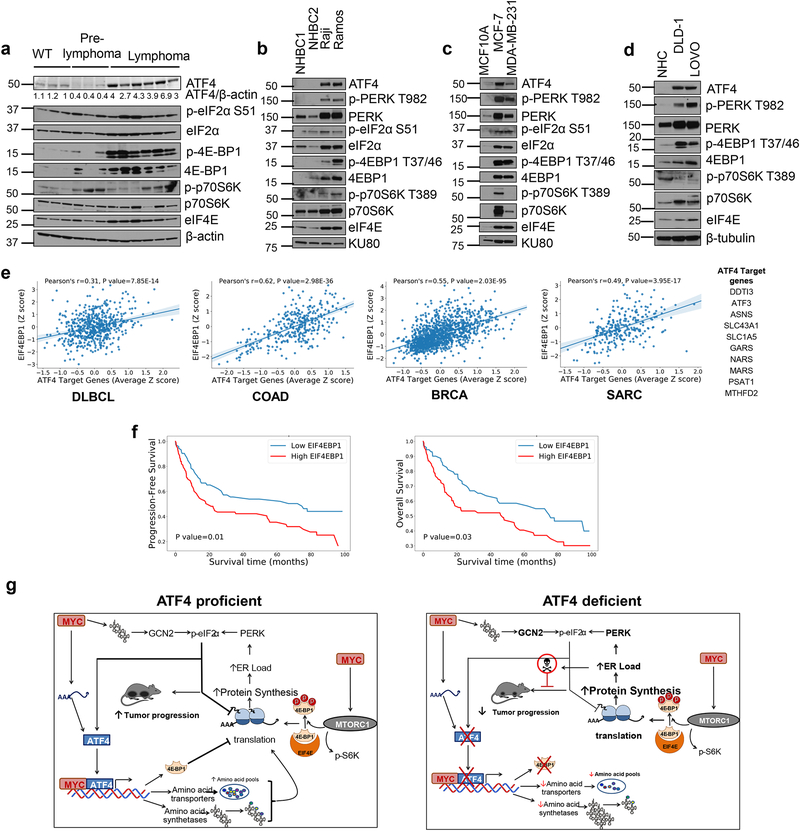
Figure 6. EIF4EBP1 positively correlates with ATF4 target gene expression and associates with poor prognosis.
a. Immunoblot of B cells isolated from WT (n=3), pre-lymphoma Eμ-myc (n=3) or lymphoma bearing Eμ-myc mice (n=6), lysates from different mice per group were used. b. Immunoblot from two normal human B cells (NHBC) and Burkitt’s lymphoma cell lines. c. Immunoblot from normal human colonocytes (NHC) or colon cancer cell lines. d. Immunoblot from MCF10 (non-tumorigenic breast epithelial cell line) or breast cancer cell lines. e. Pearson correlation between EIF4EBP1 and ATF4 target gene expression in Diffused Large B cell Lymphoma (DLBCL, n=562), Colorectal Adenocarcinoma (COAD, n=329), Breast Cancer (BRCA, n=1218) and Sarcoma (SARC, n=265). The center lines depict linear regression lines and shaded regions are 95% confidence intervals for regression lines. Datasets analyzed are listed in the methods section. Previously known ATF4 target gene list used in this analysis is shown in table. f. Kaplan-Meier plots of progression free survival (left) and overall survival (right) of DLBCL patients with high or low EIF4EBP1 expression. The survival analysis using Kaplan-Meier and two-sided log-rank test between high and low EIF4EBP1 expression groups was performed on all the patients with records of progression-free survival (left, n= 173) and overall survival (right, n=171), respectively. g. Proposed model of ATF4 and MYC co-operation in tumor progression. All immunoblots are from n=1 and unprocessed scans of blots are shown in Supplementary Fig 7.

