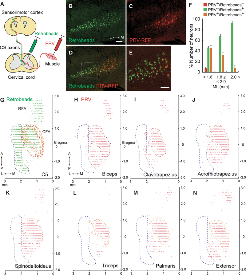
Figure 1. Retrograde Tracing Reveals Distinct Subpopulations of CSNs.
(A) Schematic diagram of the retrograde tracing of CSNs using PRVs and retrobeads, which were injected into the forelimb muscle and cervical cord, respectively. (B–E) Images of retrobeads (B) and PRV-labeled (C) CSNs in layer V of the cerebral cortex. Green, retrobeads; red, PRV-RFP. Coronal section: right, medial (M); left, lateral (L). The medial population is PRV+/retrobeads+ while the lateral population is PRV−/retrobeads+ (D). The dotted box in (D) represent the PRV+/PRV− border areas magnified in (E). Scale bars, 200 μm (B) and 100 μm (E). (F) Ratios of PRV+/retrobeads−, PRV−/retrobeads+, and PRV+/retrobeads+ neurons along the mediolateral axis of layer V. Mean ± SEM, n = 7. (G) Plotting of retrobeads+ neurons in the cortex (green dots). Top view of the cortex: PRV+ and PRV− areas determined in (H)–(N) are outlined in red-orange and blue dotted lines, respectively. RFA (AP 1.25 ± 0.05 mm to 2.60 ± 0.10 mm, ML 0.54 ± 0.04 mm to 1.42 ± 0.13 mm; n = 2); CFA (AP −0.30 ± 0.00 mm to 1.10 ± 0.00 mm, ML 0.72 ± 0.07 mm to 2.57 ± 0.02 mm; n = 2). Top, anterior (A); bottom, posterior (P); right, medial (M); left, lateral (L). (H–N) Plotting of PRV+ cortical neurons, traced from the biceps (H, proximal forelimb flexor), clavotrapezius (I, neck), acromiotrapezius (J, shoulder), spino-deltoideus (K, shoulder), triceps (L, proximal forelimb extensor), palmaris longus (M, distal forelimb flexor), and extensor carpi radialis (N, distal forelimb extensor) muscles. Note that the lateral population is PRV—/retrobeads+ (blue dotted area), whereas the medial population is PRV+/retrobeads+ (reddish orange dotted area).

