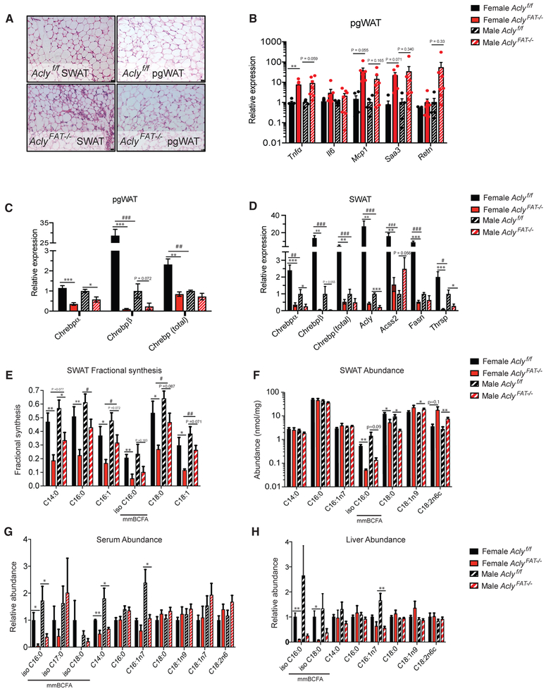
Figure 4. Females Exhibit Greater Dependence on Adipocyte ACLY for Deposition of Newly Synthesized Fatty Acids in Adipose Tissue Than Males.
(A–D) Analysis of Aclyf/f and AclyFAT−/− mice after 16 weeks on ZFD.
(A) Representative histology of female pgWAT and SWAT; scale bars represent 25 μm.
(B) Inflammatory gene expression in pgWAT by qPCR.
(C) ChREBP gene expression in pgWAT.
(D) ChREBP and target gene expression in SWAT.
(E–H) Analysis of Aclyf/f and AclyFAT−/− mice after 4 weeks on ZFD.
(E) Fractional synthesis of fatty acids in SWAT after D2O labeling of mice for 1 week.
(F) Abundance of saponified fatty acids in SWAT per mg tissue.
(G) Abundance of saponified fatty acids in serum.
(H) Abundance of saponified fatty acids in liver.
Error bars depict mean ± SEM for all panels. For all panels, n = 6 female Aclyf/f, n = 7 female AclyFAT−/−, n = 6 male Aclyf/f, n = 7 male AclyFAT−/−. Statistics by two-tailed t test; asterisks depict analysis between genotypes: *p < 0.05; **p < 0.01; ***p< 0.001; number symbols depict analysis between sexes: #p < 0.05; ##p < 0.01; ###p < 0.001. See also Figure S4.

