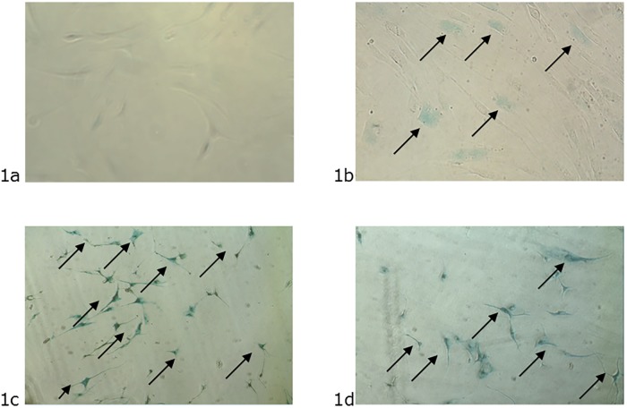
Fig 1. Images from premature senescence activity induced by stress.
1a –Primary HDF from the young donors control assay at passage 5. The absence of the blue color indicates cells with non-senescence activity. 1b –Primary HDF from elderly donors at passage 5. The blue color indicates a few senescent cells. 1c –Primary HDF from young donors submitted to UVB irradiation at passage 5. The blue color indicates cells with senescence activity. 1d –Primary HDF from young donors submitted to accelerated proliferation at passage 20. The blue color indicates cells with senescence activity.

