
Fig 1. Viral mRNA comprises 60% of the cytoplasmic mRNA at 6 hours post-infection.
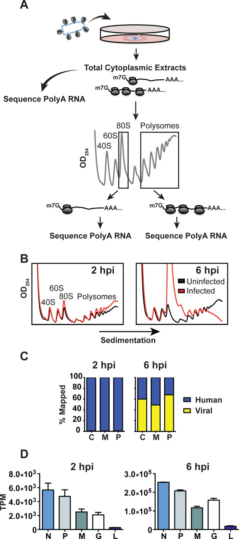
(A) Schematic of experimental design. HeLa cells were infected with VSV at a MOI of 10 and cytoplasmic extracts were prepared at 2 and 6 hpi for mRNA isolation and polysome profiling. Messenger RNA was isolated from fractions corresponding to 80S monosomes, or polysomes containing 3 or more ribosomes, and used for deep sequencing. (B) Polysome analysis of uninfected (black) or VSV (red) infected HeLa cells. Cytoplasmic extracts were sedimented through a 10–50% sucrose gradient and 0.5 ml fractions were collected while continuously monitoring absorbance at λ = 254nm. (C) Distribution of fragments mapping to the concatenated hg38 (human) and VSV genomes for cytoplasmic, monosome, and polysome samples at 2 and 6 hpi. Trimming and mapping was performed in CLC Genomics Workbench. (D) Distribution of reads among the 5 viral genes at 2 and 6 hpi. Expression level is presented as Transcripts per Kilobase Million (TPM) to normalize for gene length and library size, error bars denote standard deviation from two biological replicates.