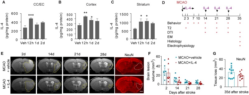
Fig 3. Intranasal delivery of IL-4 nanoparticles increases IL-4 levels in the brain but shows minimal effect on neuronal tissue loss.
IL-4 nanoparticles (50 μg/kg body weight) or an equivalent volume of vehicle (“Veh”) were applied intranasally. Brain tissues were collected at 12 h, 1 d, and 2 d after IL-4 delivery. Brain IL-4 levels were measured in the CC and EC (A), cortex (B), and striatum (C) using ELISA. n = 3–4/group. *p < 0.05, **p < 0.01, ***p < 0.001, versus vehicle-treated mice. One-way ANOVA and post hoc LSD tests. (D) Experimental design for Figs 3–7. (E) Longitudinal T2-weighted images demonstrate axial views of the same live animal brains at 2 d, 14 d, 21 d, and 28 d after MCAO. NeuN (red) immunostaining was performed 35 d after stroke. The corresponding planes of the same animals are displayed. Scale bar for NeuN images: 1 mm. (F) Brain lesion volume was assessed on T2-weighted images. n = 6/group. Two-way ANOVA followed by Bonferroni post hoc. (G) Tissue loss was measured on NeuN-stained brain slices. n = 10–11/group. Student’s t test. Data associated with this figure can be found in the supplemental data file (S1 Data). CC, corpus callosum; DTI, diffusion tensor imaging; EC, external capsule; EM, electron microscopy; IL-4, interleukin-4; LSD, least significant difference; MCAO, middle cerebral artery occlusion.

