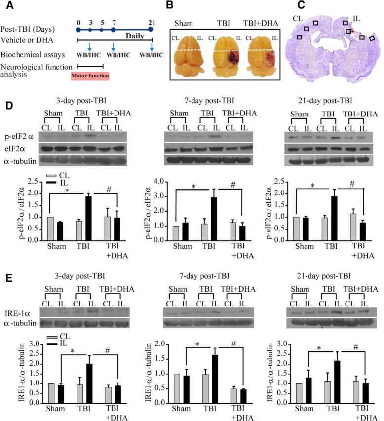
Figure 1.

Post-TBI administration of DHA reduces expression of ER stress marker protein p-eIF2α and IRE1α in the frontal cortex following TBI. A, Schematic presentation of experimental protocol. Either DMSO vehicle or DHA (16 mg/kg, i.p.) was administered at 5 min after induction of CCI, followed with the same daily dose during 3–21 d post-TBI. The time points of sample collection for Western blotting (WB) and immunohistological analysis (HI) or neuronal functional analysis were indicated. B, Representative brain images of three groups illustrates the CL and IL frontal cortex tissue (above the dotted line) harvested for immunoblotting analysis. C, Representative TBI brain section (cresyl violet stained, bregma level −1.40) illustrates sample collection in the perilesion areas (black box) of the CL and IL frontal cortex. D, Representative immunoblots of p-eIF2α and eIF2α protein expression in the CL and IL frontal cortex tissues of sham control, TBI vehicle control, and TBI+DHA animals at 3–21 d post-TBI. The same blot was probed with anti-α-tubulin antibody as a loading control. Bottom, Summary data. Data are mean ± SE; n = 4; *p < 0.05 versus sham; # p < 0.05 versus TBI. E, Representative immunoblot of IRE1α-protein expression in the CL and IL frontal cortex tissues of sham control, TBI vehicle control, and TBI+DHA animals at 3–21 d post-TBI. Data are mean ± SE; n = 4–5; *p < 0.05 versus sham; #p < 0.05 versus TBI.
