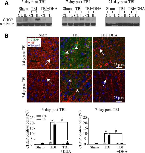
Figure 3.
DHA reduces expression of ER-resident proapoptotic protein CHOP in the frontal cortex following TBI. A, Representative immunoblot of CHOP protein expression in the CL and IL frontal cortex of three groups (see Fig. 2 legend). The same blot was probed with anti-α-tubulin antibody as a loading control. B, Confocal microscopic images showing CHOP expression in the perilesion frontal cortex tissues at 3 or 7 d post-TBI. B, Arrow, Expression of NF in neurons (red). B, Arrowhead, CHOP-positive neurons (green). Bottom, Summary data of CHOP-immunopositive cells. Values are mean ± SE (n = 4) and expressed as CHOP+ cells/100 total cells. *p < 0.05 versus CL; #p < 0.05 versus TBI.

