
Figure 7.
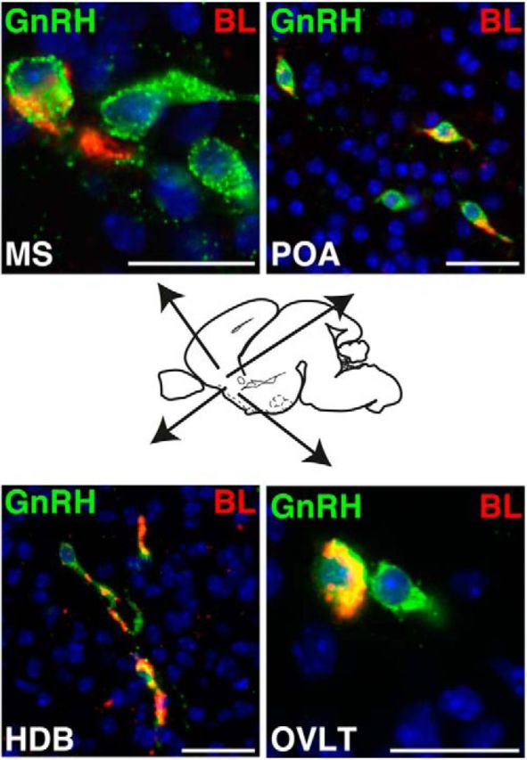
Connectivity and communication between ARC kisspeptin and GnRH neurons in utero is independent of their spatial position. Immunofluorescence for BL (red) and GnRH (green) is shown on a sagittal section through the head of a female KissIC/R26-BIZ mouse at E18.5. BL+/GnRH+ neurons are present in the medial septum (MS), the POA, the horizontal diagonal band of Broca (HDB), and the organum vasculosum of lamina terminalis (OVLT). Note that each area also contains GnRH neurons that do not communicate with kisspeptin neurons (BL−/GnRH+). Scale bars, 50 μm.