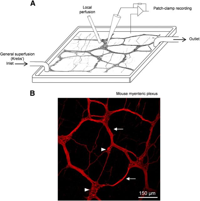
Figure 1.
In situ patch clamping of mouse myenteric neurons. A, Schematic illustration of the recording chamber for patch clamping mouse myenteric neurons maintained in situ in the LMMP preparation. The LMMP is continuously superfused with gazed Krebs' solution to provide steady oxygenation of the tissue. A local superfusion device is positioned near the selected myenteric ganglion to ensure rapid application of drugs. B, Whole-mount mouse myenteric plexus labeled with an anti-peripherin antibody. Some ganglia (arrowheads) and connective bundles (arrows) of the myenteric plexus are indicated.

