Figure 3.
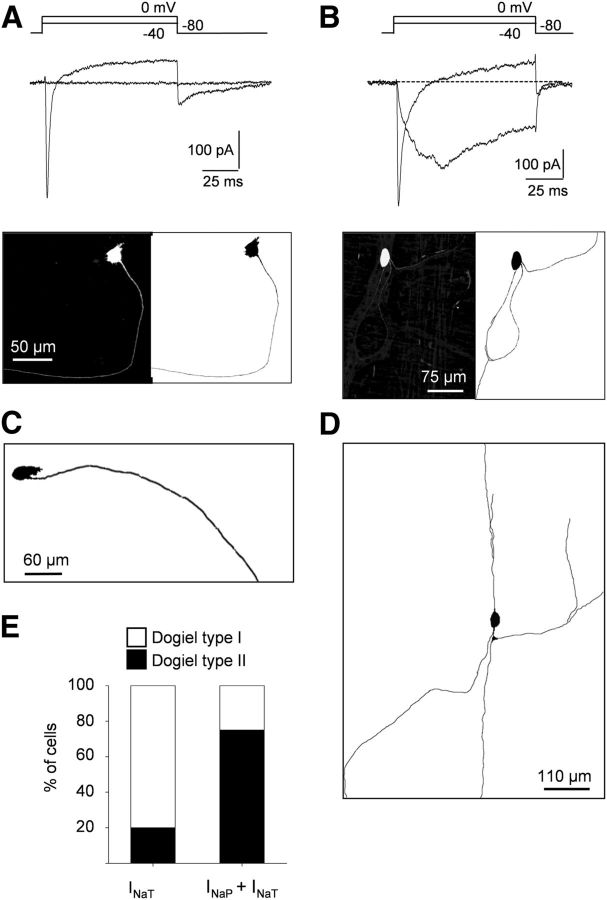
Differential distribution of INaT and INaP in myenteric neurons. A, B, Top, Na+ current traces elicited by depolarizing voltage steps in myenteric neurons exhibiting either pure INaT (A) or mixed Na+ currents composed of INaP and INaT (B). Whole-cell recordings were made with intracellular biocytin (0.2%) added to the patch pipette solution for further morphological characterization. Bottom, Morphology of neurons revealed using streptavidin–Alexa Fluor 488. Note that the neuron expressing only INaT (A) has a Dogiel type I morphology with a single long axon, whereas the neuron with prominent INaP has a Dogiel type II morphology with multiple and ramified axons (B). C, D, Examples of morphologies of myenteric neurons with pure INaT (C) or mixed Na+ current (INaP and INaT; D). E, Percentage of cells with Dogiel type I and Dogiel type II morphologies expressing either INaT or INaP + INaT.

