
Figure 4.
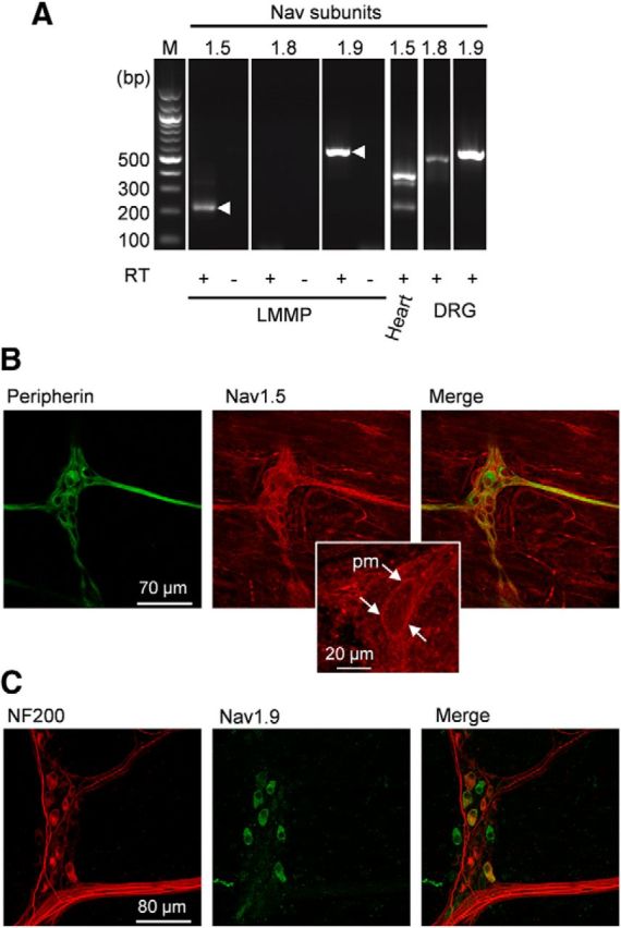
Nav1.5 and Nav1.9, but not Nav1.8, are expressed in mouse myenteric plexus. A, RT-PCR reveals transcripts for Nav1.9 and Nav1.5. cDNAs isolated from mouse LMMP, heart, and DRGs were amplified using primers to mouse Nav1.5, Nav1.8, and Nav1.9. Products of the expected sizes for Nav1.5a (220 bp) and Nav1.9 (550 bp) cDNAs were amplified from the LMMP preparation. Amplification of reverse-transcribed RNA from mouse heart and DRGs was used as a positive control. As expected, Nav1.5 (380bp) and Nav1.5a amplicons were present in heart and Nav1.8 (480 bp) and Nav1.9 amplicons were detected in DRGs. Contamination from genomic DNA was routinely tested by omitting the reverse transcriptase in the templates (RT−). M, 100 bp-ladder DNA size standard. B, The LMMP preparation was double-labeled with peripherin (green) and anti-Nav1.5 (red) antibodies. Images are projections of 59 consecutive optical sections spanning 6 μm. Middle, inset, Expanded view of a myenteric neuron with Nav1.5 plasma membrane (pm) labeling. C, The LMMP preparation was double-labeled with anti-NF200 (red) and anti-Nav1.9 (green) antibodies. Images are projections of 41 consecutive optical sections spanning 8 μm.