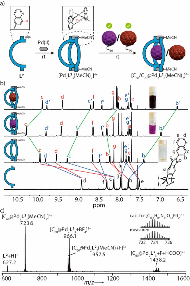
Figure 2.

Self-assembly and characterization of bowl compounds. (a) L2, comprising sterically demanding quinoline donors, reacts with PdII to the bowl-shaped host, which binds both C60 and C70. (b) 1H NMR spectra of ligand L2 (600 MHz, 298 K, CD3CN), bowl [Pd2L23(MeCN)2]4+, [C60@Pd2L23(MeCN)2]4+, [C70@Pd2L23(MeCN)2]4+ (all 0.64 mM, 298 K, CD3CN) and photos of solutions. Red and blue marked proton signals are assigned to edge and central ligands, respectively. (c) High resolution ESI mass spectrum of [C60@Pd2L23(MeCN)2]4+, prepared in pure CH3CN.
