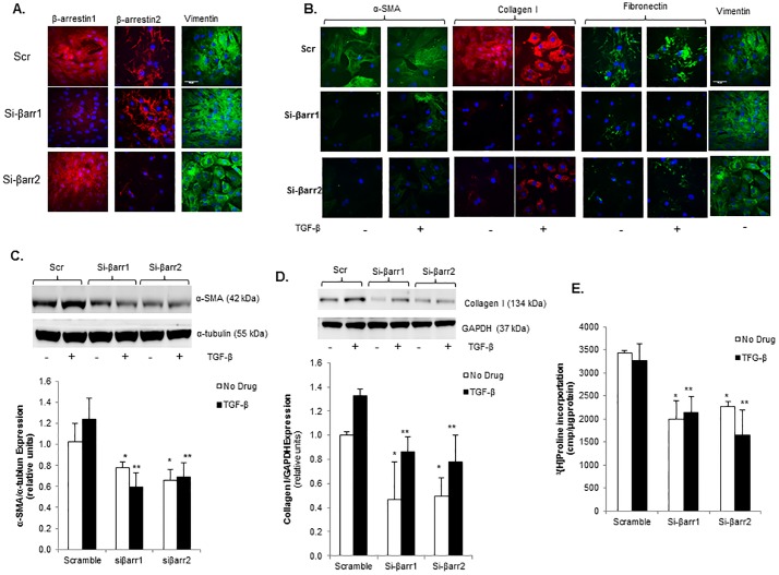
Fig 5. β-arrestin knockdown inhibits myofibroblast transformation and collagen synthesis in cardiac fibroblasts isolated post-myocardial infarction.
A. Confocal images showing β-arrestin1 and 2 stained red with Alexa Fluor 594 dye and vimentin stained green with FITC in rat cardiac fibroblasts isolated 8 weeks post-infarction treated with siRNA for β-arrestin1 (si-βarr1), β-arrestin2 (si-βarr2), or scrambled control (Scr) demonstrating successful knockdown of β-arrestins. Nuclei stained blue with DAPI. B. Confocal images showing Collagen I stained red with Alexa Fluor 594 dye and α-SMA, Fibronectin, and Vimentin stained green with FITC in 8 week post-MI CF following siRNA-mediated knockdown of β-arrestin1 and 2 treated with TGF-β vs. no drug. Nuclei stained blue with DAPI. C. Representative immunoblot (upper panel) showing decreased basal and TGF-β-stimulated α-SMA expression in 8 week post-MI CF following Si-βarr1 and Si-βarr2 treatment vs. scramble control (Scr). This membrane was stripped and re-probed for α-tubulin. Densitometric analysis shown below. *p<0.025 vs. Scr No Drug, **p<0.01 vs. Scr TGF-β; n = 4–5 in each group. D. Representative immunoblot (upper panel) showing decreased basal and TGF-β-stimulated Collagen I expression in 8 week post-MI CF following Si-βarr1 and Si-βarr2 treatment vs. scramble control (Scr). Membrane was cut based on the molecular weight and separately probed with Collagen I and GAPDH. Densitometric analysis shown below. *p<0.04 vs. Scr No Drug, **p<0.005 vs. Scr No Drug; n = 4–5 in each group. E. Collagen synthesis in 8 week post-MI CF following siRNA knockdown of β-arrestin1 and 2 under basal conditions and TGF-β stimulation. *p<0.02 vs. Scr No Drug, **p<0.02 Scr TGF-β; n = 3 in each group.

