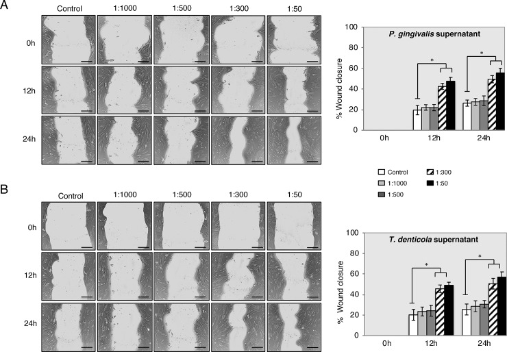
Fig 4. PDLSC migration was increased with supernatants of P. gingivalis and T. denticola by scratch wound healing assay.
Representative images are shown from 3 independent experiments and light gray area define the areas lacking cells (Scale bar 120 μm). Images were analyzed using ImageJ software to calculate wound area. Data is expressed as the mean values of percentage wound closure relative to the corresponding 0 h time point and represent the mean percentage closure ± SEM (n = 3): *p < 0.01 vs. time-matched treated control for each time-point.

