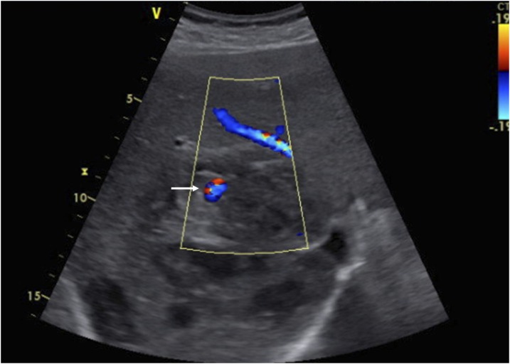
Figure 1.
Ultrasonography with color image demonstrates the hypoechoic cystic lesion in the right lobe of the liver representing a partially liquefied amebic liver abscess and an arterial aneurysm demonstrating color flow (arrow). This figure appears in color at www.ajtmh.org.

