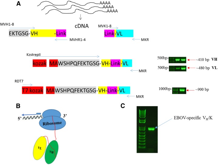
Figure 2.
(A) Schematic representation of making an scFv library from mouse spleen. (B) Schematic of the stalled antibody–ribosome–mRNA complex and position of primers used for RT-PCR recovery in the 1st cycle of ribosome display. T7 is the 5′ primer and MKR is the 3′ primer. (C) Analysis of RT-PCR recovery of VH/K cDNA in the 1st cycle. VH/K complexes were bound to EBOV GP coated to the PCR tube through 1st cycle of selection and recovery. cDNA = complementary DNA; Ebola virus = EBOV; GP = glycoprotein; RT-PCR = reverse transcription PCR. This figure appears in color at www.ajtmh.org.

