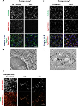
Fig. 3. Lysosomal inhibitors block mineralization.

(A and C) Confocal live imaging of 50 nM BafA-or 10 μM Vac-1–treated osteoblasts. Cells were cultured in osteogenic media containing BafA or Vac-1 and stained with Hoechst 33342 and LysoTracker Insets show higher magnification and boxed area of each channel. (B and D) SD-ADM images of BafA- or Vac-1–treated osteoblasts. Cells were cultured in osteogenic media containing BafA or Vac-1. (E) Alizain Red S staining performed without fixation. Cells were cultured in osteogenic media containing BafA or Vac-1 and stained with Alizain Red S. Representative confocal images. Scale bars, 50 μm in (A), (C), and (E); 2 μm (B) and (D).
