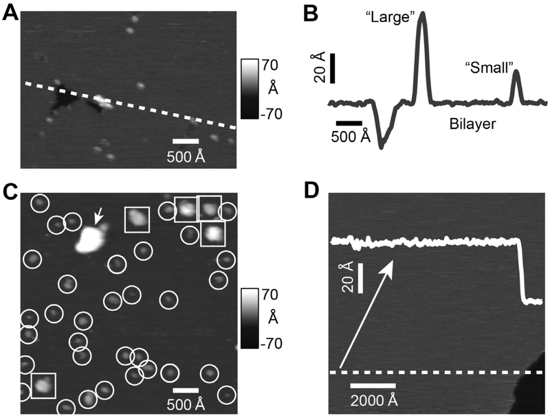
Fig. 2.
AFM images and analysis of Pgp reconstituted in liposomes. The (A) AFM image and (B) cross-section (indicated by the dashed line in panel A) shows a void as well as a “large” and a “small” protrusion. (C) A representative 1 × 1 μm AFM image showing “large” protrusions (squares), “small” protrusions (circles) and aggregates (arrow). (D) AFM image and line scan of liposomes prepared without Pgp shows no discernable features. A downward step of approximately 40 Å (bottom right) is consistent with the presence of bilayer.

