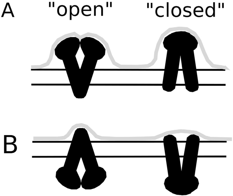
Fig. 7.

Model of Pgp conformational dynamics in a lipid bilayer. The protrusion envelope (gray line) produced from the (A) C and (B) EC domains of Pgp (black) with cartoons of the “open” (left) and “closed” (right) Pgp structure. The two horizontal lines roughly reflect the membrane position deduced using the OPM or TMDET algorithms.
