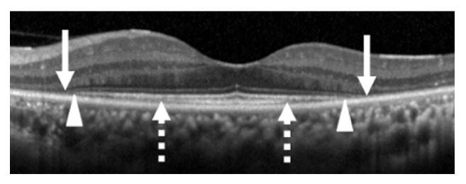
Figure 2.

Optical coherence tomographic image centered on the fovea of a patient with RP. Arrows indicate the ends of the external limiting membrane line, arrowheads indicate the ends of the ellipsoid zone line, and dashed arrows indicate the end of the interdigitation zone.
