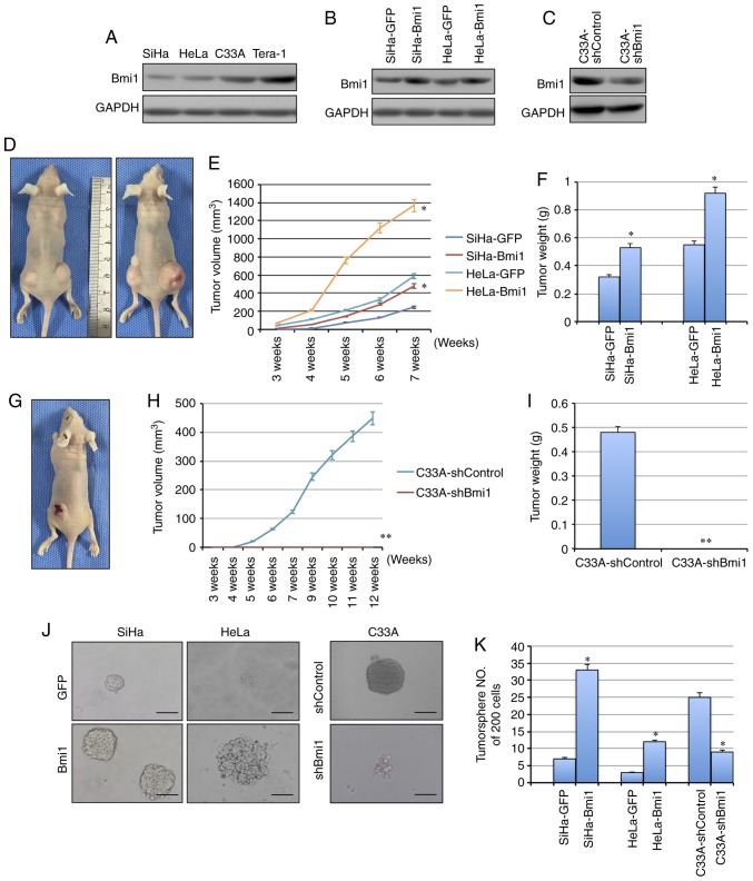
Figure 2.
Bmi1 promotes carcinogenicity and tumor sphere formation ability in cervical cancer. (A) The expression of Bmi1 protein in three cervical cancer cell lines was detected by western blotting; the teratoma cell line Tera-1 was used as the positive control. (B) Bmi1 protein expression was evaluated in Bmi1-overexpressing SiHa and HeLa cells by western blotting. (C) Bmi1 protein expression was evaluated in Bmi1-silenced C33A cells by western blotting. (D) Tumor growth was assessed over (E) 7 weeks, as well as the (F) the tumor net weight of nude mice inoculated with SiHa-Bmi1, SiHa-GFP, HeLa-Bmi1 and HeLa-GFP cells with three replicates (1×10⁵ cells). Additionally, (G) tumor growth was measured over (H) 12 weeks, as well as the (I) tumor weights of nude mice inoculated with C33A-shBmi1 and C33A-shControl cells with three replicates (1×10⁶ cells). (J) The tumor sphere formation assay was performed in FBS-free medium in low-density cultures per 200 cells with six replicates, and then (K) the number of tumor spheres formed by Bmi1-overexpressing SiHa and HeLa cells and Bmi1-silenced C33A cells was calculated. Bars=SE. *P<0.05, **P<0.01 vs. respective control. Bmi1, polycomb complex protein Bmi1; GFP, green fluorescent protein; sh, short hairpin.

