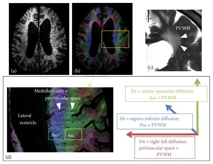
Figure 2.
(a) Susceptibility-weighted image (SWI) shows the medullary veins running in the periventricular white matter. (b) Fusion of SWI and color-coded FA map. The periventricular white matter is divided into two regions; medial blue and lateral green areas, which correspond to the projection and association fibers, respectively. (c) In cases with ventriculomegaly, PVWH is often observed on T2-weighted image. (d) The medullary vein runs through the association and projection fiber areas (Pro and Asc). We hypothesized Dx reflects perivascular space of the deep white matter vessels and PVWH. Therefore, Dx partially include the glymphatic system. Dy and Dx are supposed to reflect the association fiber plus PVWH and the projection fiber plus PVWH, respectively.

