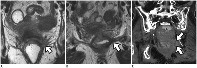
Fig. 3. Distal rectal cancer with left levator ani muscle involvement.
A. On baseline MRI, ulceroinfiltrative mass is observed in distal rectum with tumor infiltration into left levator ani muscle (arrow). B. On post-CRT MRI, signal drop is seen within entire tumor. However, shrunken tumor still invades levator muscle (arrow). Traditional APE was performed. Histopathology revealed tumors with pathologic tumor regression grade 3 and positive pCRM (not shown). C. Follow-up CT 6 months after operation shows recurrent mass at remnant left levator muscle (arrows). CRT = chemoradiotherapy, MRI = magnetic resonance imaging

