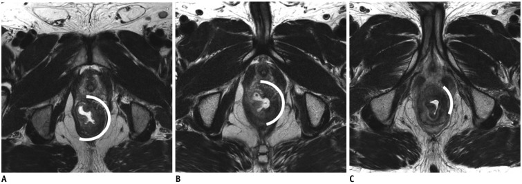
Fig. 4. Progressive tumoricidal effect and fibrotic change in rectal cancer after routine timing window.
A. On baseline MRI, semi-circular mass with intermediate SI is noted at left lateral wall of rectum (curved line indicates circumferential tumor extent). B. Post-CRT MRI obtained at four weeks after completion of CRT shows increased proportion of fibrotic change as well as decreased proportion of tumor SI, indicating tumor regression. Patient refused surgical option at that time. C. On post-CRT MRI obtained at 22 weeks, proportion of fibrotic change is further increased with decreased circumferential tumoral extent. Histopathology after subsequent APE revealed ypT2 (not shown). SI= signal intensity

