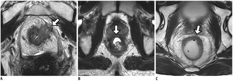
Fig. 8. Tumor penetration through MRF with MRF thickening which suggests high risk for positive pCRM even after completion of CRT.
A. Post-CRT MRI shows circumferential mass with intermediate SI. Tumor penetration to MRF with MRF thickening (arrow) is noted. Pathologic result after TME was positive pCRM (not shown). B. In another patient, post-CRT MRI still demonstrates tumor infiltration anteriorly into prostatic gland (arrow). After APE with partial prostatectomy, pCRM was found to be positive. C. On post-CRT MRI in another patient, tumor with anterior MRF threatening by spicules of treated tumor (arrow) is noted. However, MRF thickening or penetration is not demonstrated (arrow). Pathologic result after TME was negative pCRM (not shown).

